Pseudoaneurysm Repair

Pseudoaneurysm Repair

A pseudoaneurysm, also known as a false aneurysm, is a vascular condition characterized by a breach in the arterial wall leading to blood collection outside the vessel, contained by the surrounding tissues. Unlike a true aneurysm, which involves dilation of all three layers of the arterial wall, a pseudoaneurysm results from a disruption in the vessel wall integrity, causing blood to pool and form a pulsatile hematoma.

Pseudoaneurysms are clinically significant because they can lead to complications such as rupture, compression of adjacent structures, thrombosis, or distal embolization. Timely diagnosis and appropriate management, including pseudoaneurysm repair, are crucial to prevent morbidity and mortality. This article provides a detailed, reader-friendly guide on pseudoaneurysm repair, covering anatomy, causes, symptoms, diagnosis, treatment options, procedural details, postoperative care, risks, prognosis, and when to seek medical attention.

Anatomy / Background

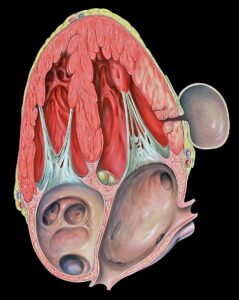
To understand pseudoaneurysms, it is essential to review the basic anatomy of arteries. Arteries consist of three layers:

  • Intima: The innermost endothelial lining.
  • Media: The middle muscular layer providing structural support.
  • Adventitia: The outer connective tissue layer.

A true aneurysm involves dilation of all three layers, whereas a pseudoaneurysm occurs when there is a disruption in the intima and media, allowing blood to escape into the surrounding tissue but contained by the adventitia or perivascular tissue. This creates a sac that communicates with the arterial lumen through a neck.

Common sites for pseudoaneurysms include arteries subjected to trauma or invasive procedures, such as the femoral artery (after catheterization), radial artery, and carotid artery.

Causes / Etiology

Pseudoaneurysms can arise from various causes, including:

Symptoms / Clinical Presentation

Patients with pseudoaneurysms may present with:

Diagnosis

Diagnosis involves a combination of clinical examination and imaging studies:

  • Physical Examination: Identification of a pulsatile mass with a bruit or thrill.
  • Ultrasound Doppler: The first-line, non-invasive imaging modality that visualizes the pseudoaneurysm sac, neck, and blood flow pattern (to-and-fro flow).
  • Computed Tomography Angiography (CTA): Provides detailed vascular anatomy, especially for deep or complex pseudoaneurysms.
  • Magnetic Resonance Angiography (MRA): Alternative imaging for patients with contraindications to contrast.
  • Conventional Angiography: Used for both diagnosis and therapeutic intervention.

Treatment Options

Treatment depends on the size, location, symptoms, and risk of complications.

Non-Surgical Management

  • Ultrasound-Guided Compression Repair (UGCR): Manual compression of the pseudoaneurysm neck to induce thrombosis; suitable for small pseudoaneurysms.
  • Ultrasound-Guided Thrombin Injection: Injection of thrombin directly into the pseudoaneurysm sac to promote clotting; highly effective and minimally invasive.
  • Observation: Small, asymptomatic pseudoaneurysms may be monitored for spontaneous resolution.

Surgical Treatment

  • Open Surgical Repair: Indicated for large, expanding, infected, or complicated pseudoaneurysms. Involves excision of the pseudoaneurysm and arterial repair or bypass.
  • Endovascular Repair: Placement of covered stents to exclude the pseudoaneurysm from circulation; minimally invasive and preferred in select cases.

Procedure Details (Surgical Repair)

Open Surgical Repair

Open surgical repair typically involves the following steps:

    1. Anesthesia: General or regional anesthesia is administered.
    2. Incision: A surgical incision is made over the affected artery.
    3. Exposure: The artery and pseudoaneurysm sac are carefully exposed.
    4. Control of Blood Flow: Proximal and distal arterial control is achieved using vascular clamps.
    5. Excision: The pseudoaneurysm sac is opened and removed.
    6. Arterial Repair: The arterial defect is repaired by direct suture, patch angioplasty, or interposition grafting depending on the size of the defect.
    7. Hemostasis and Closure: After ensuring no bleeding, the wound is closed in layers.
    8. Postoperative Monitoring: The limb is monitored for perfusion and complications.

Postoperative Care / Rehabilitation

  • Monitoring: Vital signs, limb perfusion, and wound site are closely observed.
  • Pain Management: Analgesics are administered as needed.
  • Antibiotics: Given if infection is suspected or confirmed.
  • Physical Activity: Gradual mobilization is encouraged to prevent complications.
  • Follow-Up Imaging: Ultrasound or CTA to assess repair integrity.
  • Lifestyle Modifications: Control of risk factors such as hypertension and smoking cessation.

Risks and Complications

  • Potential risks and complications include:

    • Bleeding or Hematoma Formation
    • Infection
    • Nerve Injury
    • Thrombosis or Embolism
    • Recurrence of Pseudoaneurysm
    • Arterial Occlusion or Stenosis
    • Wound Healing Problems

Prognosis

With timely and appropriate treatment, the prognosis for pseudoaneurysm repair is generally excellent. Minimally invasive techniques have improved outcomes and reduced recovery times. However, untreated pseudoaneurysms can lead to serious complications, including rupture and limb ischemia.

When to See a Doctor

Seek medical attention if you experience:

  • A new or enlarging pulsatile mass near an artery.
  • Persistent pain or swelling after arterial catheterization or trauma.
  • Signs of limb ischemia such as numbness, coldness, or color changes.
  • Sudden onset of neurological symptoms near the affected area.
  • Fever or signs of infection near a vascular access site.

Conclusion

Pseudoaneurysm repair is a critical intervention to manage a potentially life-threatening vascular condition. Understanding the anatomy, causes, symptoms, and treatment options empowers patients and caregivers to recognize the condition early and seek appropriate care. Advances in minimally invasive techniques have enhanced treatment efficacy and safety. If you suspect a pseudoaneurysm or have risk factors, consult a healthcare professional promptly for evaluation and management. Early diagnosis and treatment are key to preventing complications and ensuring optimal outcomes.

Welcome to
Dr Sahaja Vascular Surgeon

Aenean porta orci nam commodo felis hac ridiculus fusce fames maximus erat sed dictumst blandit arcu suspendisse sollicitudin luctus in nec

Make an Appointment.